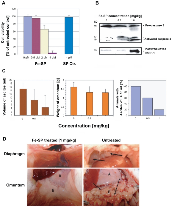
Figure 5.
Chemotherapeutic effect of Fe-SP in an animal ovarian cancer cell model. (A) Effect of Fe-SP on the viability of rat ovarian cancer cells (NUTU-19) in vitro. Rat ovarian cancer cells (NUTU-19) were plated into 96 well flat bottom plates and treated with various concentrations (0.5–4 μM) of Fe-SP or 4 μM noncomplexed SP. The MTS viability assay was carried out as described (Materials and Methods). (B) In vivo activation of Caspase-3 and in-activation of PARP-1 in ovarian cancer cell-derived tumors after Fe-SP treatment. NUTU-19 derived tumor tissue of nontreated or Fe-SP treated (0.5 mg/kg or 1.0 mg/kg bodyweight) rats was pooled for each treatment group. Cell lysates were prepared in the presence of a broad range of proteinase inhibitors to prevent protein degradation and the expression of caspase-3 and PARP-1 was analyzed by immunoblotting (see Material and methods). Cell lysates 50 μg of total cellular protein/lane were separated on a 12% SDS-polyacrylamide gel. PARP-1 was visualized using a primary antibody solely recognizing the inactivated/cleaved protein. For caspase-3 an antibody which recognizes the full length pro-form as well as activated/cleaved fragments of the protein was used. Thus, a direct conversion of the precursor into activated caspase-3 was monitored for all samples allowing direct comparison of loading between samples from treated and nontreated animals. (C) Volume of ascites and weight of omentum from Fe-SP treated rats with SKOV-3 derived tumors. Graphs show a trend for decreased ascites and omental tumor burden after Fe-SP (0.5, 1.0 mg/kg) treatment. (D) Image of diaphragm and omentum. Example of complete response of a SKOV-3 derived tumor to Fe-SP at concentration of 1 mg/kg body weight. Following treatment both the diaphragm and omentum are disease free.

