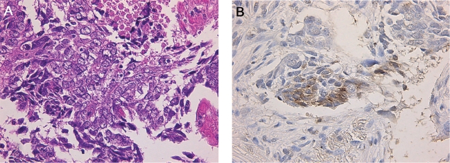
Figure 3.
Histologic and immunohistochemical analysis of the tumor specimen obtained during a computed tomography-guided percutaneous tumor biopsy. A: individual tumor cells were polygonal in shape with relatively abundant cytoplasm and vesicular nucleus, and were arranged in nests with area of coagulative necrosis. B: Tumor cells

