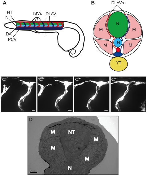
Figure 1. Preliminary studies of anastomosis in zebrafish.
(A) Longitudinal diagram of 48 hpf zebrafish embryo, with neural tube (NT, green), notochord (N, turquoise), posterior cardinal vein (PCV, dark blue), dorsal aorta (DA, red), intersegmental vessels (ISVs, dark blue and red according to origin) and dorsal lateral anastomotic vessel (DLAV) shown in the trunk/tail region. (B) Cross section of zebrafish trunk; muscle blocks (M, pink) and yolk tube (YT, yellow) are also shown. (C) In vivo live confocal imaging of the anastomotic process showing two adjacent ISVs sprouting from the dorsal aorta and extending filopodia which eventually fuse to form the DLAV. (D) Ultrathin TEM sections give a general overview of the fish anatomy but it is not possible to identify the ISVs or DLAV as they do not have the lumen characteristic of blood vessels at this point. Bar (C–C′′′) 10 µm, (D) 10 µm.

