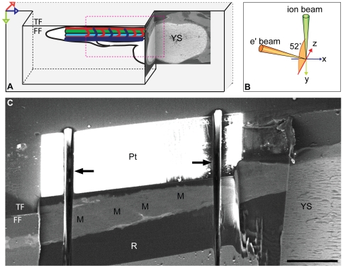
Figure 2. Sample preparation for FIB/SEM of zebrafish.
(A) Diagram showing orientation of the zebrafish after resin embedding and trimming with a diamond knife. The AOI in the zebrafish tail is placed in an overhang of resin to optimise milling and mimise curtaining artefacts. The head and yolk sac (YS) are used as markers for locating the AOI, and are exposed during trimming, seen here in BSE images from the FIB/SEM. Top face (TF), front face (FF). Arrows represent x (blue), y (green) and z (red) axes. (B) Orientation of the electron and ion beams with respect to the front face (FF) of the block. (C) Platinum (Pt) was deposited onto the top face (TF) of the block to stabilise the edge during milling. The sample was coarse milled with the gallium ion beam to expose muscle blocks (M) at the predicted area of interest (AOI) prior to the milling run. Trenches (arrows) were then milled on either side of the AOI to reduce artifacts due to redeposition of sputtered material on the blockface. Resin (R). Bar (C) 100 µm.

