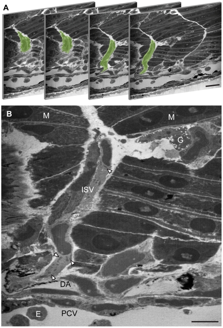
Figure 3. Localisation of the intersegmental vessels by FIB/SEM.
(A) FIB/SEM sections of a 32 hpf zebrafish embryo trunk with the ISV highlighted in green. Slices are non-consecutive sections 661, 669, 687 and 691 where one slice is 50 nm thick. (B) The pericardinal vein (PCV) and dorsal aorta (DA) can be observed as lumenized vessels containing nucleated erythrocytes (E). The ISV sprouts from the DA and migrates in the space between the muscle block (M) and midline structures. Subcellular structure can be resolved including mitochondria (arrows) and dense granules (G) . Bar (A) 30 µm, (B) 10 µm.

