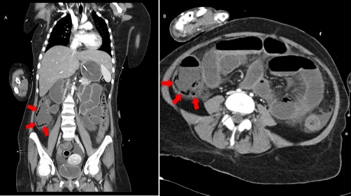
Figure 1A and B.
Abdominal and thoracic computer tomography of patient 1 coronal view: Multiple air filled cysts in the intestine wall along the right hemicolon can be seen (red arrows). Diagnosis Pneumatosis cystoides intestinalis was made. The sonographic findings (free abdominal air near the portal vein) were not confirmed in CT.

