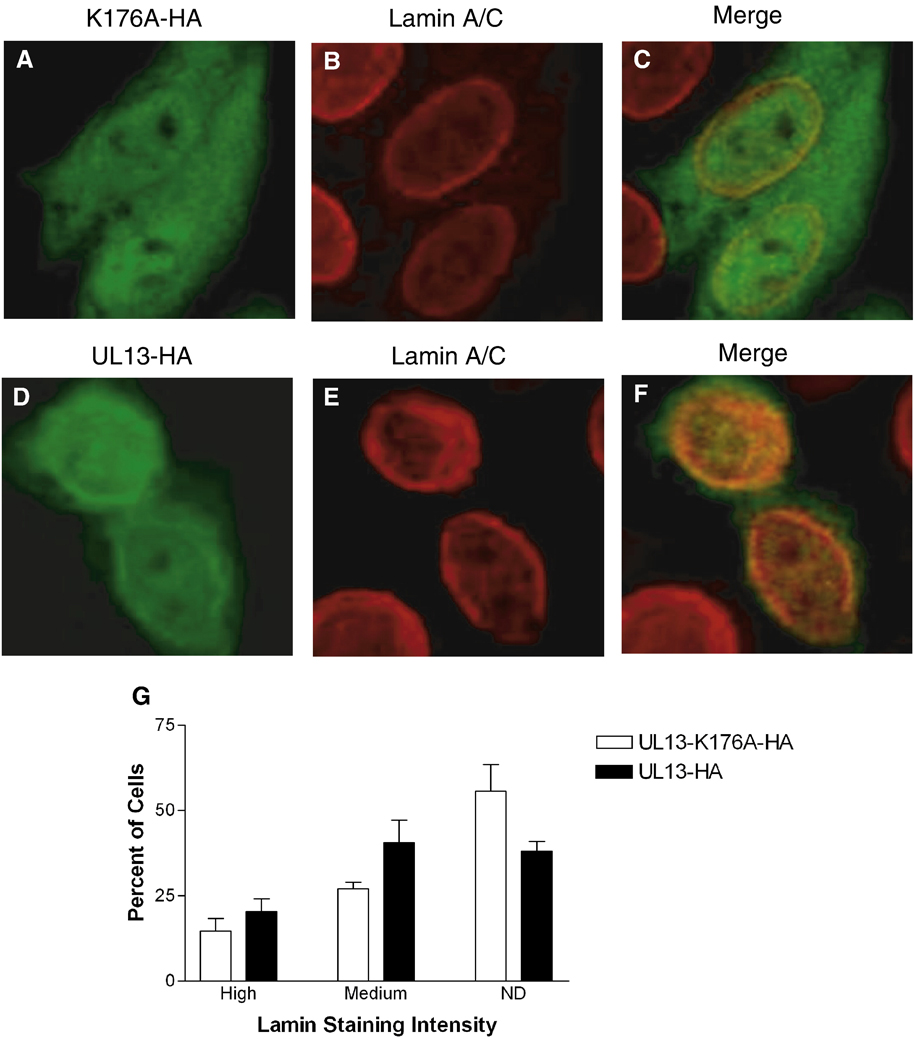
Figure 2. Lamin A and C staining with a polyclonal antibody does not differ in HeLa cells expressing UL13-HA or UL13-K176A-HA.
HeLa cells were transfected with UL13-K176A-HA (A–C) or UL13-HA (D–F) expression constructs. At 18 h post-transfection, cells were fixed, stained with anti-HA antibody (green) and a polyclonal lamin A/C antibody (red), and examined by confocal microscopy. Images showing both transfected and untransfected cells were merged (C and F). All images were obtained at the same microscope settings and representative images are shown. Ten random fields were analyzed and lamin staining in each cell was ranked as high or medium intensity or not detectable (ND) by a masked observer. (G) Graph shows the mean ± SD from three experiments. No significant differences between UL13-K176A-HA and UL13-HA were observed (P = 0.2482 to 0.4685).

