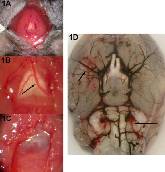
Fig. 1.
Procedure for mouse model of SAH. (A) Initial exposure consists of midline suboccipital incision and lateral retraction of strap muscles. (B) A closer view of the underlying transparent atlanto-occipital membrane with the visible subarachnoid vein (arrow). (C) After transection of the vein, blood quickly fills the subarachnoid space. The muscles and skin are closed on one layer and the animals are recovered. (D) After 24 h, there is evidence of blood in the basilar cisterns (arrows). The blood vessels are casted with India ink.

