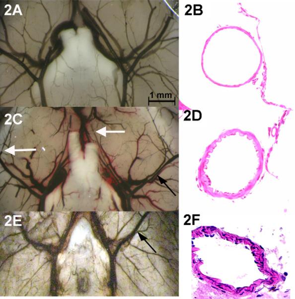
Fig. 2.
Evidence of CVs in an animal with SAH (A) compared to control (D). Note the segmental narrowing of the MCA (closed arrows) and the abrupt interruption of dye in medium vessels (open arrows). Vessels are casted with India ink. H&E preparation of meninges in control (B) shows normal blood vessel wall morphology. In SAH (E) the arterial smooth muscle layer of the MCA artery is thickened and the wall shows rogations consistent to the histology seen in human CVs. When compared to a model of arterial blood injection into the subarachnoid space, the degree of vasospasm by angiography was similar (C) and the arterial thickening was similar although more prominent in the smaller pial vessels than in the MCA (F).

