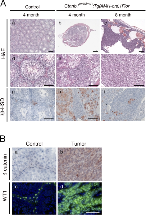
FIG. 2.
Tumor progression in Ctnnb1tm1Mmt/+;Tg(AMH-cre)1Flor male mice. Aa–Af) Normal histology in testes of 4-mo-old control males (low magnification, Aa; high magnification, Ad), and progressive development of testicular tumors in Ctnnb1tm1Mmt/+;Tg(AMH-cre)1Flor males of different ages (low magnification, Ab and Ac; high magnification, Ae and Af), shown by hematoxylin and eosin staining. Ag–Ai) Immunostaining with Leydig cell marker 3β-HSD identified Leydig cells in testes of Ctnnb1tm1Mmt/+;Tg(AMH-cre)1Flor and control mice at different ages. B) β-Catenin (red) and WT1 (green) immunostaining on control testes and tumors. Tumor cells were positive for β-catenin and WT1 staining, indicating their Sertoli cell origin (Bb and Bd, respectively). Bar = 250 μm (Aa–Ac) and 100 μm (Ad–Bd).

