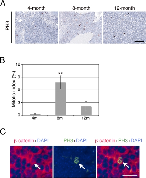
FIG. 3.
Increased cell proliferation in tumor cells of Ctnnb1tm1Mmt/+;Tg(AMH-cre)1Flor mice at 8 mo of age. A) Immunostaining with the proliferation marker PH3 identified mitotic cells in testes of Ctnnb1tm1Mmt/+;Tg(AMH-cre)1Flor mice at different ages. The number of mitotic cells increased significantly at 8 mo of age, which is consistent with the tumor onset time. B) Quantitative analysis showed that approximately 8% of cells were positive for PH3 staining at 8 mo of age, as compared with less than 1% and approximately 2% at 4 mo of age and 1 yr of age, respectively. **P < 0.01. C) Double-immunofluorescent staining of β-catenin and PH3 on tumors at 8 mo of age. Left, β-catenin (red) was highly accumulated in tumor cells; middle, one PH3-positive (green) proliferating cell; right, merge showing the proliferating cell was positive for β-catenin staining (arrow). Bar = 100 μm.

