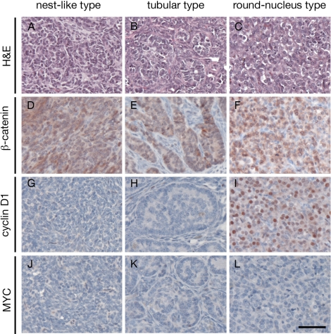
FIG. 4.
Microscopic features of testicular tumors in β-catenin stabilized mutants. A–C) Three types of tumor morphologies developed in mutant mice: tumor cells forming a nest-like pattern as the dominant type, tumor cells forming a tubular pattern, and tumor cells with very round nuclei. D–F) Immunohistochemical analysis of β-catenin on tumor samples. β-Catenin staining were observed in all three types of tumor cells. G–I) Immunohistochemical analysis of cyclin D1 on tumor samples. In tumors with a nest-like pattern and tubular pattern, very few tumor cells were positive for cyclin D1 staining. However, in the round-nucleus tumor type, almost all tumor cells were positive for cyclin D1 staining. J–L) Immunohistochemical analysis of MYC showed that tumor cells were negative for MYC staining in all three types of tumor cells. Bar = 50 μm.

