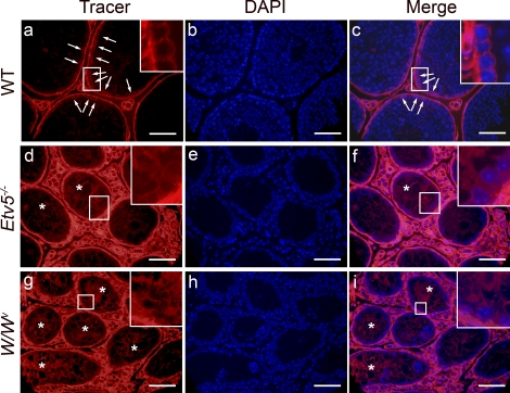
FIG. 6.
Biotin tracer localization in adult WT, Etv5−/−, and W/Wv mice. A biotin tracer was injected into testicular interstitial space of adult mice and was allowed to penetrate the tissue for 30 min. b, e, and h) Cell nuclei are visualized with DAPI stain, and merged images of tracer and DAPI show the penetration of tracer into the seminiferous epithelium and lumen. Enlargements are shown of areas outlined by white boxes. a–c) In WT mice, the tracer stopped at the level of the BTB (arrows). Tracer is seen surrounding only cells lining the basement membrane. d–f) In Etv5−/− mice, tracer is observed surrounding all cells and along strands of Sertoli cell membranes (*) and reaching the tubule lumen. g–i) In W/Wv mice, biotin tracer surrounds all cell types and extends into the lumen along Sertoli cell membranes (*). Bar = 50 μm.

