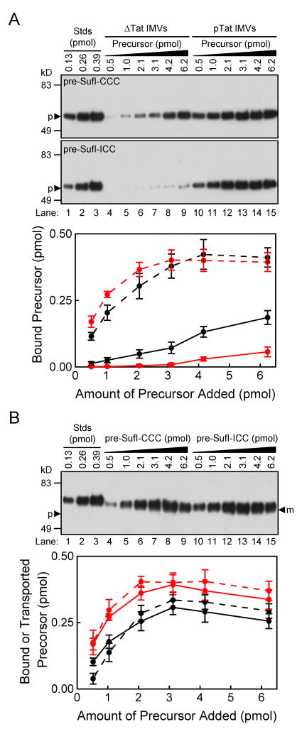
Figure 5. Membrane binding and transport efficiency of pre-SufI-ICC.
All gels in this figure are anti-SufI immunoblots. (A) Concentration dependence of membrane binding efficiency. The amount of pre-SufI-CCC (black) and pre-SufI-ICC (red) bound to ΔTat (solid) and pTat IMVs (dashed) was quantified (n = 3). (B) Correlation between transport efficiency and the amount of translocon-bound precursor. The gel shows the concentration dependent transport efficiency of pre-SufI-CCC and pre-SufI-ICC using 4 mM NADH. The plot shows the amount of transported pre-SufI-CCC (black) and pre-SufI-ICC (red) from three independent experiments (solid lines). The amount of translocon-bound protein was calculated from the data in A as described in Fig. 1C (dashed lines).

