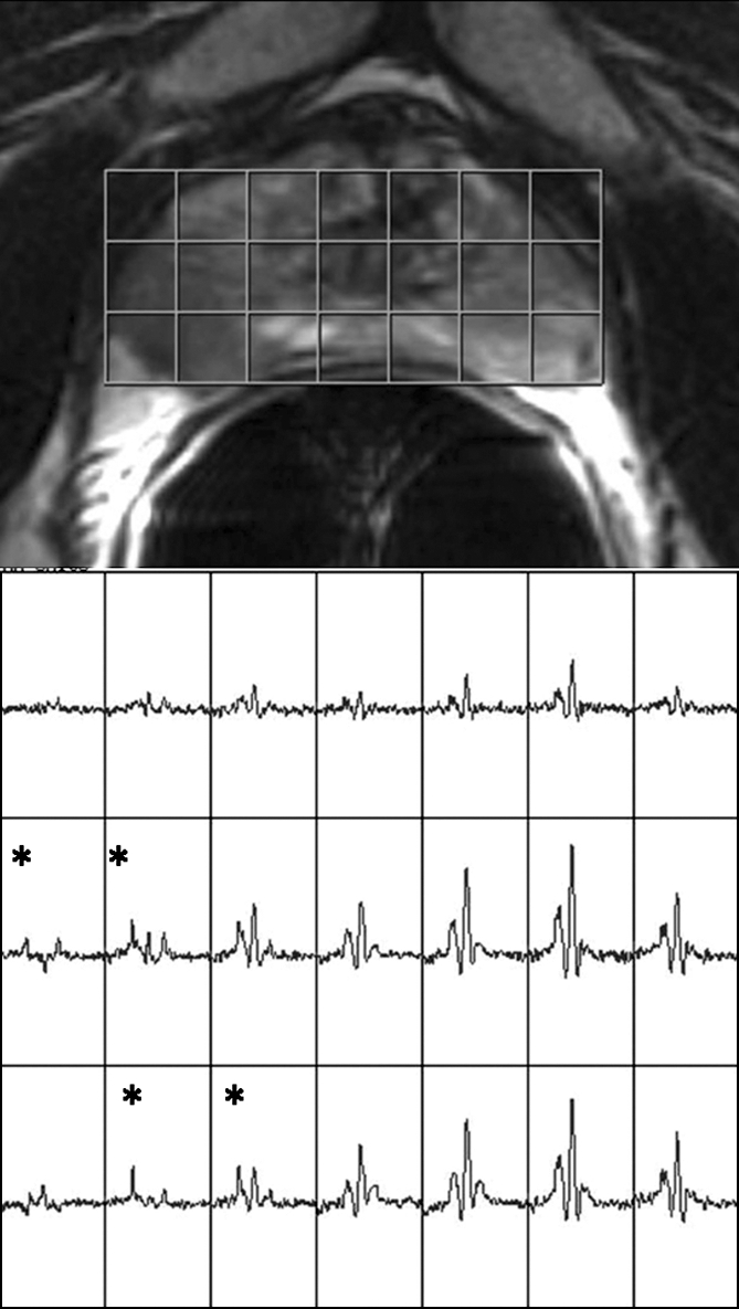
Figure 4b:

Clinical stage T1c prostate cancer in 60-year-old man. (a) Transverse T2-weighted endorectal MR image (4016/105, echo train length of 10, three acquired signals, 14-cm field of view, 256 × 192 matrix, 3-mm section thickness, no intersection gap) of prostate shows focal low-signal-intensity tumor (white arrow) in peripheral zone in right middle portion of prostate gland. The tumor is associated with capsular bulging, which is consistent with extracapsular extension (black arrow), which was later confirmed at surgical-pathologic analysis. (b) MR spectroscopic grid overlaid on image in a. Asterisks indicate suspicious voxels (elevated choline level and undetectable or reduced citrate level) in right posterior peripheral zone. MR spectroscopic imaging parameters are the same as those for Figure 3.
