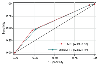
Figure 5a:
Graphs illustrate accuracy in diagnosing clinically nonimportant prostate cancer with endorectal MR imaging or combined endorectal MR imaging–MR spectroscopic imaging (MRSI) in patients with clinical stage T1c disease. (a) Reader 1 had an AUC of 0.63 (95% CI: 0.55, 0.71) with use of MR imaging alone and 0.62 (95% CI: 0.54, 0.70) with use of combined MR imaging–MR spectroscopic imaging. (b) Reader 2 had an AUC of 0.72 (95% CI: 0.64, 0.80) with use of MR imaging alone and 0.71 (95% CI: 0.63, 0.79) with use of combined MR imaging–MR spectroscopic imaging.

