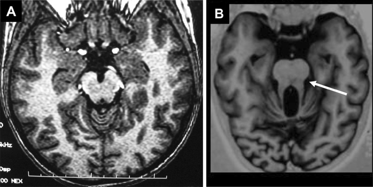
Fig. 3.
Molar tooth sign on brain magnetic resonance imaging (MRI). Brain MRI axial image at the level of the superior cerebellar peduncles of a control subject (a) and an affected individual (b) showing abnormally increased depth of the interpeduncular fossa, narrowing of the midbrain tegmentum, and thickening of the superior cerebellar peduncles, all of which contribute to the radiologic feature known as the molar tooth sign (white arrow)

