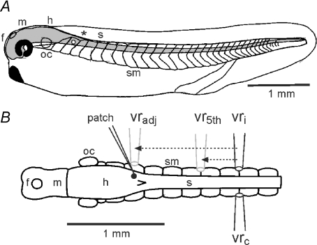
Figure 1. The hatchling Xenopus tadpole and adjustments to timing measurements according to recording sites.
A, diagram of the tadpole in side view to show the CNS (grey) and its regions (f, m, h: fore-, mid- and hind-brain; s: spinal cord; oc: otic capsule;*: obex) with swimming muscles (sm) in chevron shaped segments. White dot indicates the neuron in B. B, diagram of the CNS and swimming muscles in dorsal view to show typical sites for electrodes to record from a single neuron (patch) and ventral roots on the ipsilateral (vri) or contralateral (vrc) side. Measured timings of neuron activity were adjusted as though relative to recordings from either an assumed, adjacent ventral root (vradj) or the 5th post-otic ventral root (vr5th) on the same side as the recorded neuron (arrows and grey electrodes; see text for details).

