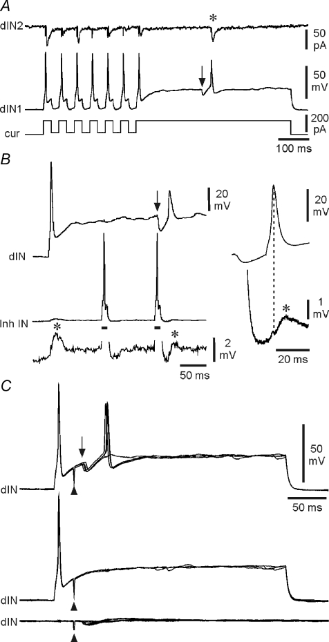
Figure 8. Post-inhibitory rebound in dINs.
A, rebound spikes to current pulses (dIN1), then a rebound spike following a spontaneous IPSP (at arrow), producing an EPSC (*) in a postsynaptic neuron (dIN2). B, when held just above spike threshold, a dIN fires on rebound following an IPSP (at arrow) elicited by the second of two spikes evoked in an inhibitory interneuron (Inh IN) by depolarising current (bars). Evoked and rebound spikes in the dIN both evoke EPSPs (*) in the inhibitory interneuron. Right, averages of 5 rebound spikes in the dIN and resulting EPSPs in the inhibitory interneuron. C, rebound in a dIN, depolarised just above spike threshold, following IPSPs (arrow) evoked by stimulation (at arrowhead) of the contralateral side of the spinal cord (upper overlapped traces). No rebound occurs where IPSPs fail (middle traces) or when the dIN is at rest (lower traces).

