
Figure 2.
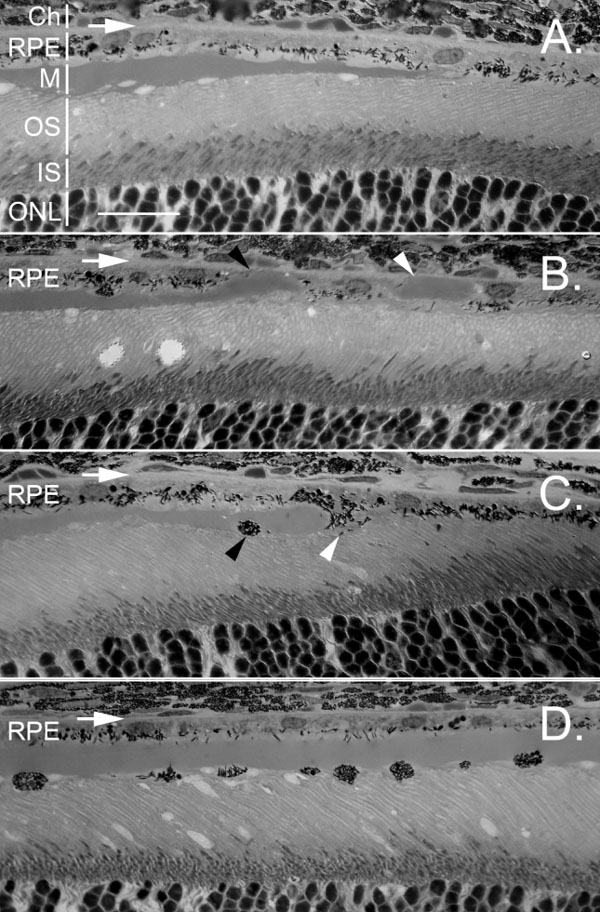
Retinal pigment epithelium cells mobilization in response to subretinal Matrigel. The position of Bruch's membrane is indicated by a horizontal white arrow in each panel. A: The edge of Matrigel layer in the subretinal space is shown 1 day after injection. No retinal pigment epithelium (RPE) cells mobilization is detected. RPE mobilization was detected 5 days after injection of Matrigel (B-D). In B, a small piece of gel (white arrowhead) had been relocated to the sub-RPE space. On the left side of the same retinal section, two RPE cells formed a gap, entrapping the tip of the main gel body (black arrowhead). The gel tip was thus in direct contact with Bruch's membrane. C: A cell at the edge of the gel (white arrowhead) extended between photoreceptors and the gel. Another had migrated to the photoreceptor side of the gel (black arrowhead). D: In the central region of the Matrigel layer, many cells had migrated to the photoreceptor side. Ch indicates choroid; M indicates Matrigel layer; IS indicates inner segments; ONL indicates outer nuclear layer. The scale bar represents 20 μm.