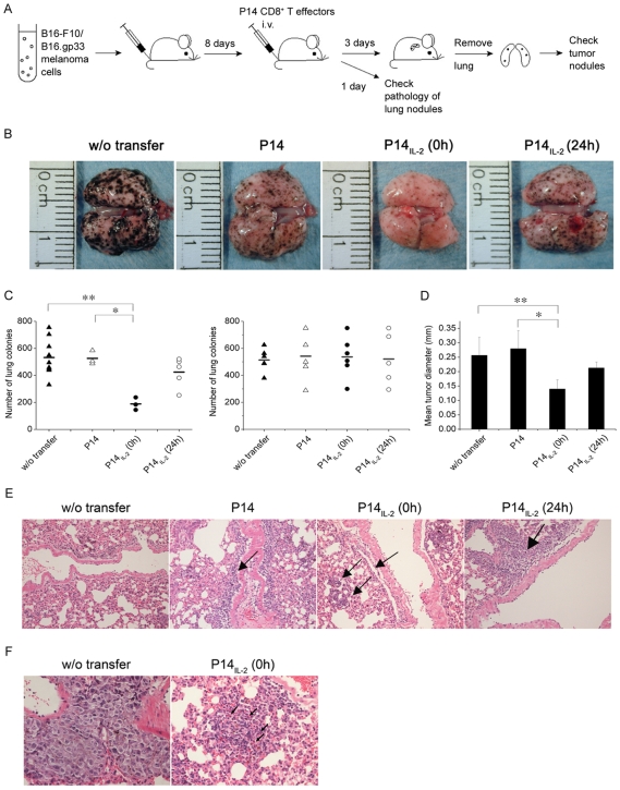
Figure 6. IL-2 signal in vitro at CD8+ T-cell priming may enhance better tumor-eradicating efficacy in vivo.
(A) B16-F10 or B16.gp33 melanoma cells were inoculated intravenously into host mice (0.5×10⁶ cells per mouse), followed by injection of P14 effector T cells (1×10⁷ cells per mouse) at 8 days after the tumor injection. The mice were sacrificed one day later for pathology study of lung nodules, or three days later for checking lung tumor nodules. (B) Three days after P14 effector cells transfer, the lungs of two mice in each group was shown. (C) Three days after P14 effector cells transfer, the number of B16.gp33 (left) and B16-F10 (right) melanoma colonies in lung was recorded. (D) Three days after P14 effector cells transfer, the size of B16.gp33 melanoma colonies in lung was recorded. (E and F) One day after P14 effector cells transfer, the lung was fixed in formalin, followed by H& E staining. The pathology of lung nodules was visualized by microscopy at 100× (E) and 400× (F) of magnification. w/o transfer, without cell transfer. P14, transfer with P14 CD8+ T effector cells in absence of IL-2 at priming, P14IL-2 (0 h) and P14IL-2 (24 h) in presence of IL-2 at 0 hour and 24 hours after stimulation, respectively. Similar results were obtained in three independent experiments. *, **, significantly statistical difference by student t- test, p<0.05. Arrow in (E), lymphocytes. Arrow in (F), apoptotic body.

