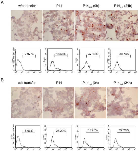
Figure 7. IL-2 signal at priming drove better anti-tumor CTL function in vivo.
(A) Expression of granzyme B in tumor-infiltrating lymphocytes. Upper panel. At 4 hours after effector cells transfer, the lung tissue was subjected to immunohistochemical staining by FITC anti-granzyme B antibody (1 µg/mL), followed by anti-Fluorescein peroxidase. After wash, the tissue was subjected to NOVA-RED and Hematoxylin staining. Lower panel. At 4 hours after transfer of CFSE-labeled effector cells, the lymphocytes in the lung were isolated and subjected to intracellular staining by PE anti-granzyme B antibody, and analysis by flow cytometry. (B) Expression of IFNγ in tumor-infiltrating lymphocytes. Upper panel. At 4 hours after effector cells transfer, the IFNγ production in the lung was checked by immunohistochemical staining by anti-IFNγ antibody (5 µg/mL), followed by biotin goat anti-rat Ig (1 µg/mL) and streptavidin-peroxidase (1∶1000). After wash, the tissue was subjected to NOVA-RED and Hematoxylin staining. Lower panel. As (A), the production of IFNγ was also checked in the CFSE-labeled effector cells using intracellular staining by PE anti-IFNγ antibody, and analysis by flow cytometry. Similar results were obtained in three independent experiments.

