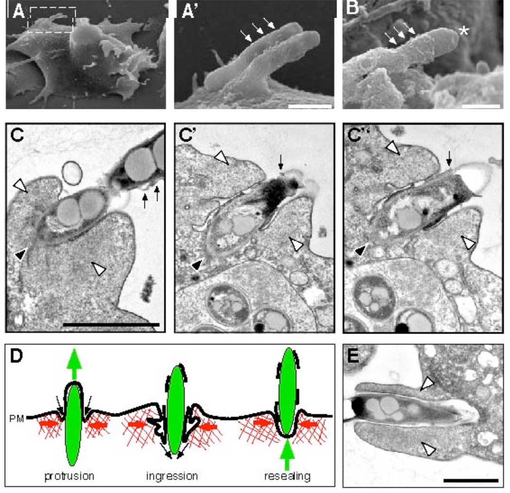
Fig. 3.
Electron microscopy of ejecting bacteria and schematic representation of an ejection event. Scanning electron microscopy (A–B, A’ is the magnified inset of A) showed bulges of the plasma membrane (small arrows), some ruptured at the tip of the ejecting bacterium (B, asterisk). Scale bars 0.5 µm. (C) Serial sections through an ejectosome revealed the organization of the Factin (white arrowheads) and the plasma membrane. The posterior of the ejecting bacterium (black arrowhead) was in the cytosol. The bacterium was separated from the F-actin by the invaginated plasma membrane, which was tightly apposed to its surface. Membrane fragments were scattered along the extracellular part of the bacterium (small arrows). (D) Schematic representation of an ejection. During outward movement of a bacterium (green), the F-actin barrel exerts contraction (red arrows), and the invaginating plasma membrane (PM) reseals at the posterior of the bacterium (black arrows), maintaining a tight septum despite partial membrane rupture. (E) Micrograph of a phagocytic cup. The actin-filled lamellipodia (white arrowheads) extend and engulf a bacterium. Scale bars 1µm.

