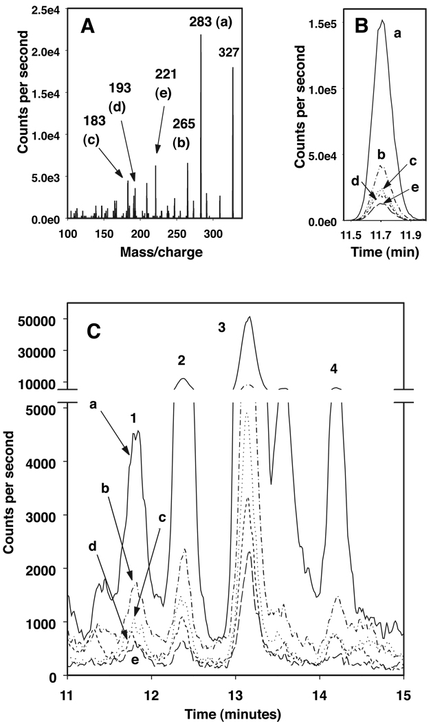
Figure 2. LC-MS identification of 2,3-dinor-PGF1α.
(A) Fragmentation pattern of 2,3-dinor-PGF1α standard, m/z 327 to 283 (a), 265 (b), 183 (c), 193 (d) or 221 (e).
(B) LC-MS chromatogram of 2,3-dinor-PGF1α standard showing tracings of each of the fragments (a–e) identified in A.
(C) LC-MS chromatogram of a urine extract with tracings of peaks identified with SRM profiles showing each of the fragments (a–e, identified in A) that matched those of 2,3-dinor-PGF1α (2,3-dinor-F1, retention time same as the standard, labeled 1; F1-12, labeled 2; F1-13, labeled 3, and F1-14 labeled 4).

