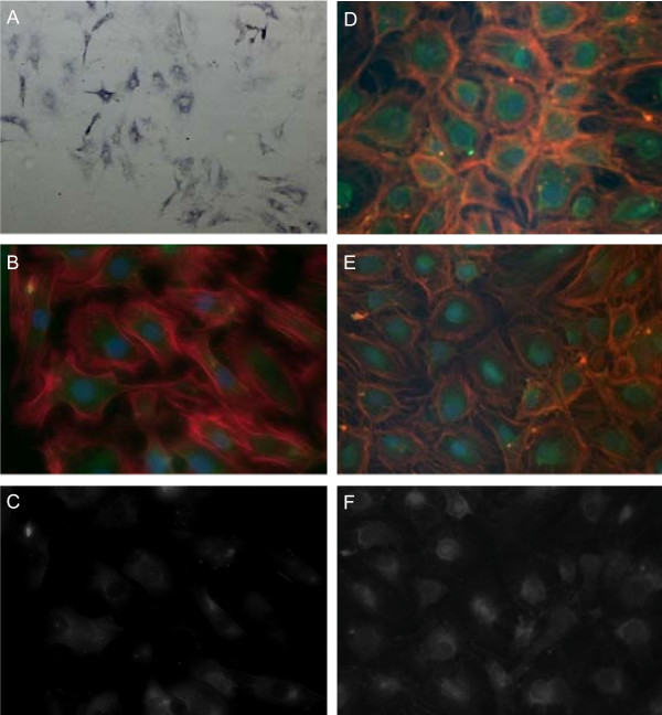
Figure 1.
Immunostainings of cBAL111 cells in vitro. The cells were stained using antibodies against GS (A) (blue, 40× magnification, 2 days in culture), albumin (B, C) (green, 400× magnification, 2 days in culture) and CK19 (D) or CK18 (E, F) (both green, 400× magnification, 15 days in culture). In the CK18, CK19 and albumin staining, cells were further visualized by phalloidin, binding to actin (red), and DAPI, binding to the nuclei (blue) (B, D, E). Corresponding single stains of albumin and CK18 are shown in C and F.

