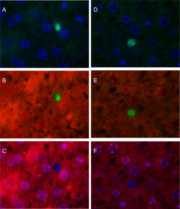
Figure 4.
FISH analysis (40× magnification) of mouse livers after transplantation of GFP marked cBAL111 cells. The probe hybridizing with human DNA was visualized using a FITC labeled antibody (A, B, D and E), showing green nuclei, the probe hybridizing with murine DNA was visualized using a Texas Red labeled antibody (red nuclear staining on top of cytoplasmic background autofluorescence, B, C, E and F). All nuclei were counterstained using DAPI (blue A, C, D and F). Picture sets A-C and D-F both show a human cell that is negative for the murine DNA.

