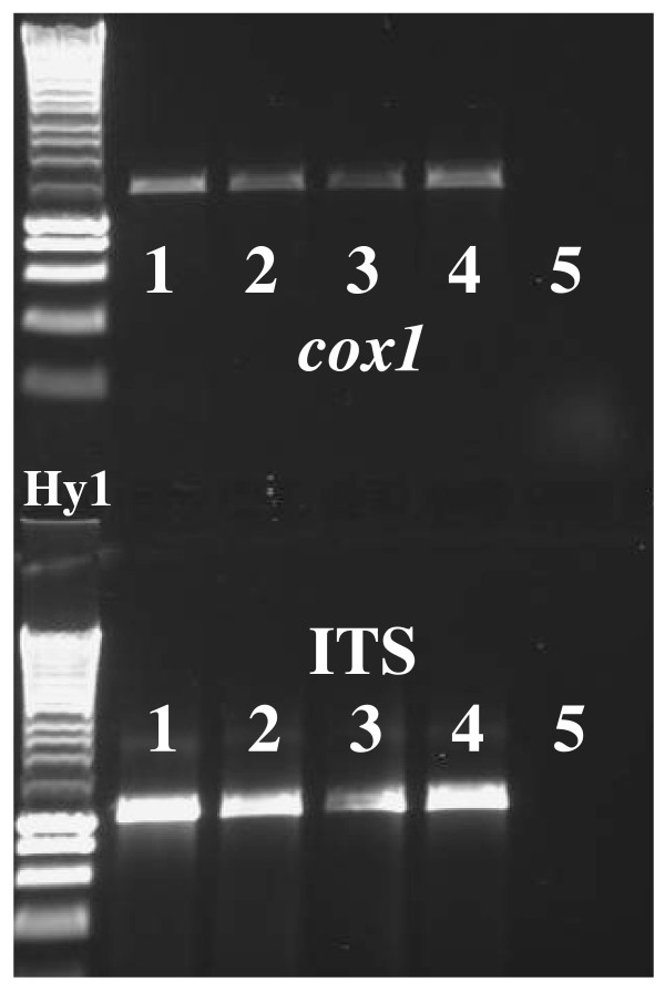
Figure 2.
Gel image showing 3 μl of PCR amplicons amplified from DNA extracts of individual schistosome larval stages and eggs, preserved in RNAlater®. The image was captured using the UVP gel doc system. Tracks (Hy1 = Hyperladder 1) 1. RNAlater® preserved adult worm (Positive control). 2. RNAlater® preserved miracidia. 3. RNAlater® preserved cercaria. 4. RNAlater® preserved egg. 5. (Negative control) 2 μl of the water that the miracidia were hatched in was mixed with 5 μl of RNAlater®. The DNA extraction protocol was carried out on this mix and the resulting elute used in the PCR.

