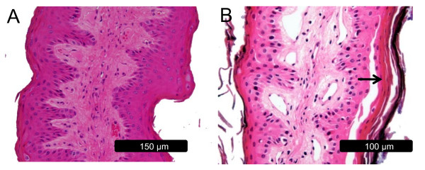
Figure 3.
Light micrographs of rumen papillae biopsied during the high forage and high grain diets. A: rumen papillae from the high forage diet with an intact stratum corneum and granulosum. B: rumen papillae from the high grain diet displaying sloughing of the stratum corneum and demarcation of cells through the epithelial layers (arrow).

