Fig. 3.
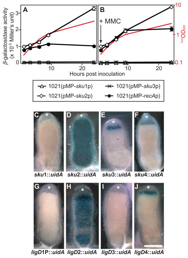
Expression of S. meliloti NHEJ genes in free-living cells and bacteroids. Panels A and B represent the levels of β-galactosidase activity (given in 103 Miller’s units) for the promoter region of sku1, sku2, sku3 and recA fused to lacZ respectively. Assays were performed at 1, 3, 6, 9 and 24 h after subculture. In B, 0.2 μg ml−1 of MMC was added to induce SOS response. The values reported represent the means of three independent experiments with standard errors (error bars). Growth curves of a representative strain (monitored by 1cmOD600) were also shown (red curves). Panels C–J represent histochemical localization of β-glucuronidase activity in nodule hand-sections. Nodules were harvested from alfalfa plants infected with Rm1021 strains carrying NHEJ gene–uidA fusions (Rm1021sku1::pJH104, Rm1021sku2::pJH104, Rm1021sku3::pJH104, Rm1021sku4::pJH104, Rm1021ligD1P::pJH104, Rm1021ligD2::pJH104, Rm1021ligD3::pJH104 and Rm1021ligD4::pJH104; C–J respectively). β-Glucuronidase activity was visualized as blue precipitates of the chromogenic substrate 5-bromo-4-chloro-3-indolyl glucuronide (X-Gluc). A total of 30–50 nodules from five plants were examined for each fusion. The meristematic zone of nodule is marked with white asterisks. Scale bar represents 0.5 mm.

