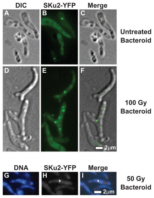
Fig. 5.
Subcellular localization of SKu2 in bacteroids. Panels A–I represent subcellular localization of SKu2–YFP fusion proteins in S. meliloti bacteroids by epi-fluorescence/DIC microscopy. Alfalfa plants infected with Rm1021sku2-yfpmut2 were irradiated by 100 Gy of IR (D–F), 50 Gy of IR (G–I) or mock-treated (A–C). Nodules were immediately harvested and crushed. Long, blanched bacteroid cells were visually distinguished from non-differentiated S. meliloti cells or plant-derived materials (A and D). In contrast to the free-living cells, the membrane of bacteroids could not be stained by FM4-64 (data not shown). Localization of SKu2-YFP was visualized in bacteroids as represented in green (B and E) or white (H). Cellular DNA was stained by DAPI as represented in blue (G). Epi-fluorescence views of different filters and DIC views were merged to analyse relative localization (C, F and I). No focus was observed in bacteroid cells of the Rm1021 wild-type strain (data not shown). Scale bar represents 2 μm.

