
Figure 3.
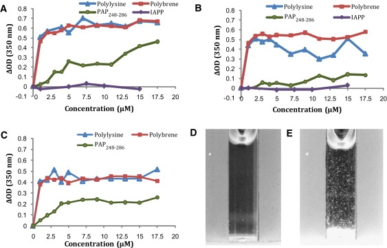
Lipid aggregation of liposomes of varying composition detected by changes in turbidity at pH 4: (A) 7:3 POPC/POPG, (B) viral membrane composition, and (C) host cell membrane composition. Reported values are the changes in the absorbance at 350 nm relative to the control immediately after addition of the peptide or polymer and vigorous mixing. (D) A photograph of the sample containing 100 nm 7:3 POPC/POPG liposomes at 500 μM concentration before the addition of 17.5 μM PAP248-286. (E) A photograph of the same sample 10 min after the addition of 17.5 μM PAP248-286.