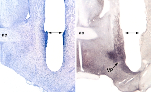
Fig. 1.
Probe placement in a Nissl-stained section of a dialyzed rat (left) and in a substance P immunostained section (right). Dark substance P staining clearly outlines the VP (right). From the base of the guide cannula tracts (double headed arrows), the dialysis probes extend ~2 mm, with the ventralmost 1 mm containing the dialysis membrane (0.6 mm OD). Substance P staining and the presence of the decussating anterior commissure (ac) were used initially to determine the appropriate AP coordinates for probe placement in SD (−0.30) and LE (+0.20) rats. Other coordinates (from Bregma) were the same for both strains: ML ± 2.5 mm; DV –6.3 mm (after Paxinos and Watson 1998)

