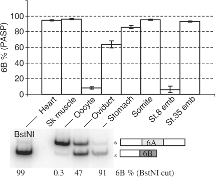
Figure 4.
PASP of α-tropomyosin in Xenopus embryos and organs. RNAs were extracted from oocytes, stage 8 or 35 embryos, dissected embryonic somites or the indicated adult organs, and α-tropomyosin-splicing patterns were analysed by PASP. The percentages of exon 6B (mean ± SD of six independent RNA preparations) are shown (upper panel). Adult heart, oviduct stomach, and oocyte RNA were also analysed by radioactive RT–PCR as in Figure 2B (lower panel). The percentages of 6B exon (restricted by BstNI) are indicated under the gel.

