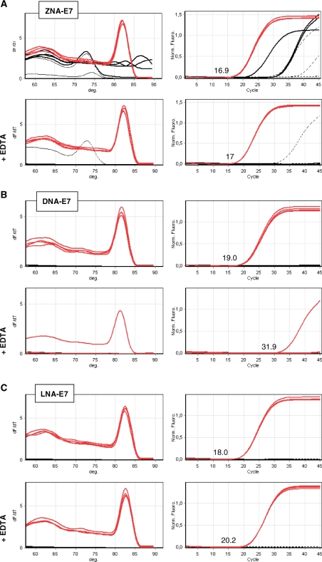
Figure 2.
SYBR Green-based qPCR of HPV16-E7 using ZNA, DNA and LNA primers. Target genomic DNA (10 ng, red lines), control DNA (10 ng, black thin lines) and no template control (black dotted lines) were amplified in quadruplicate with 100 nM of ZNA-E7 (A), DNA-E7 (B), LNA-E7 (C) primer pairs. All reactions were performed using Sensimix NoRef DNA kit (Quantace) without (upper panels in A–C) and with 1 mM EDTA (lower panels in A–C) as indicated. Melting curves (left panels) and amplification plots (right panels) are represented. Cq values for target amplification are indicated. Cycling profile was 95°C (10 s), 60°C (1 min).

