Figure 1.
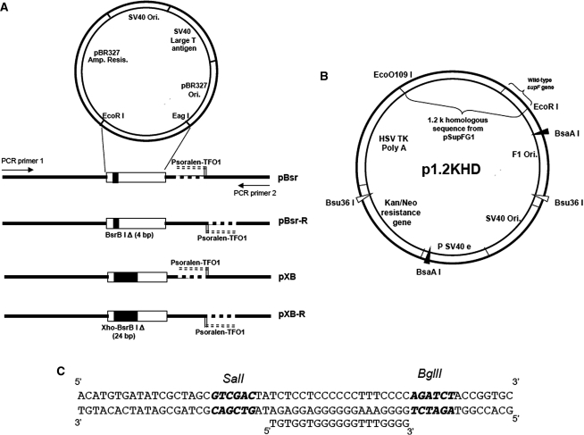
Schematic of recipient (A) and donor (B) plasmids. (A) Psoralen ICL sites are at the same position in all plasmids, the dashed line indicates the purine-rich strand bound by pTFO1. This TFO binding sequence was inserted in the plasmids via an insertion of 73 bp shown in (C). (B) The donor plasmid, p1.2kHD, has a 1.2 kb fragment homologous to the recipient plasmids, but contains the wild-type supF gene. p1.2kHD has two restriction enzyme sites for BsaAI and Bsu36I, which are absent from the recipient plasmids. (C) The insertion sequence containing the TFO1 binding sequence. The unique SalI site is used to confirm the presence of the ICL, and the unique BglII site is used to confirm the presence of the triplex structure formed by TFO binding.

