
Figure 1.
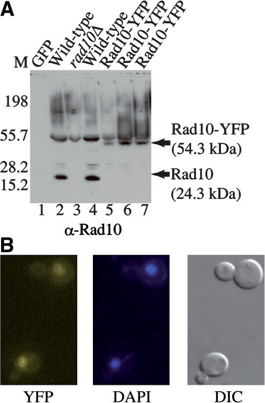
(A) The Rad10-YFP strain exhibits an increased polypeptide size consistent with Rad10 expressed as a YFP fusion protein. Immunoblot of WCE from the indicated yeast strains probed with a Rad10 antibody. 1 µg total protein was loaded to each lane. Lane 1: GFP protein (Santa Cruz Biotechnology); lane 2: SX46a WILD-TYPE; lane 3: BJRad10Δ; lane 4: W303-1A wild-type (W1588-4C); lanes 5 and 7: Rad10-YFP (WPF006-4C); lane 6: Rad10-YFP Rad52-CFP (WPF019-26C). The wild-type strains (lanes 2 and 4) show a 24 kDa Rad10 band while the Rad10-YFP strains (lanes 5–7) show a 54 kDa Rad10-YFP band. A rad10 deletion mutant (rad10Δ, lane 3) shows only background. Markers were run on the left. (B) The Rad10-YFP protein exhibits nuclear localization. YFP fluorescence (left panel) colocalizes with the DAPI fluorescence signal (center panel) confirming the nuclear localization of the YFP fluorescence. A DIC image of the same cells is also shown (right panel).