Figure 2.
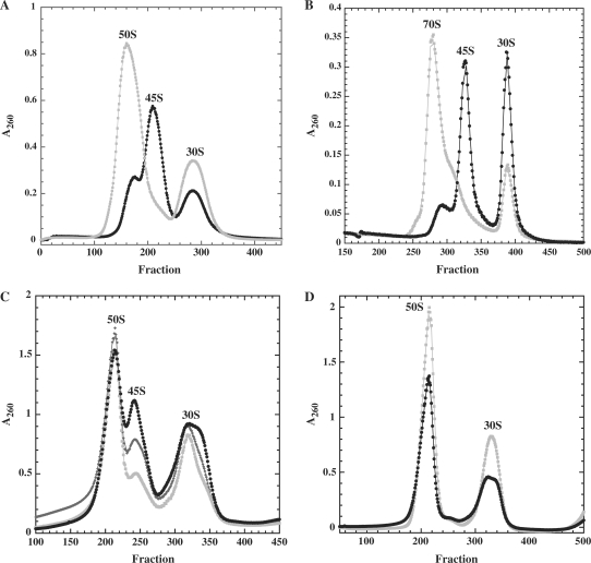
(A) DbpA R331A 50S subunits (black circles) and 70S ribosomes (grey squares) collected from gradients containing 10 mM Mg2+ analyzed on a second gradient containing 1 mM Mg2+. (B) DbpA R331A 50S (grey circles) and R331A 45S (black circles) incubated with native 30S subunits and analyzed on gradients containing 10 mM Mg2+. (C) Ribosomes from cells overexpressing DbpA mutants, DbpA R331A (black circles), DbpAK53A (dark grey diamonds) or DbpA E154A (grey squares) were analyzed on sucrose gradients containing 1 mM Mg2+. (D) BL21(DE3) (grey squares) and BL21(DE3) ΔdbpA (black circles) were grown to midlog at 22°C and analyzed on sucrose gradients containing 1 mM Mg2+.

