Figure 2.
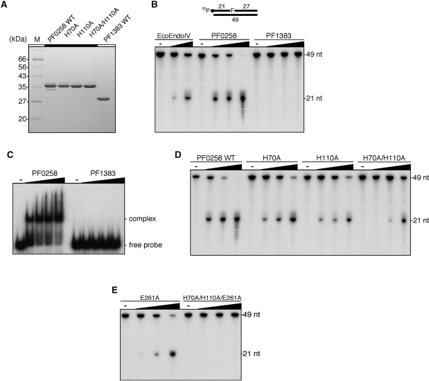
Identification of AP endonuclease activity. (A) Purified WT putative endonuclease IV proteins from P. furiosus and their mutant proteins (2 µg) were analyzed by 12.5% SDS–PAGE. The gel was stained with Coomassie Brilliant Blue. (B) An AP site-containing DNA substrate (100 fmol) was incubated with increasing amounts of the putative endonuclease IV proteins from P. furiosus (0, 5, 25 and 125 nM), as described in the ‘Materials and Methods’ section. Escherichia coli endonuclease IV (New England Biolabs Inc.) (0, 5 and 25 units) was used as a positive control of the reaction. The reaction mixtures were analyzed by denaturing PAGE, and the DNA was visualized by autoradiography. (C) An AP site-containing DNA substrate (100 fmol) was incubated with increasing amounts of the putative endonuclease IV proteins from P. furiosus (0, 50, 100, 150 and 200 nM), as described in the ‘Materials and Methods’ section. The protein–DNA complexes were analyzed by 6% PAGE, followed by autoradiography. (D) and (E) An AP site-containing DNA substrate (100 fmol) was incubated with increasing amounts of WT PF0258 protein and its mutants (0, 5, 25 and 125 nM), as described in the ‘Materials and Methods’ section.

