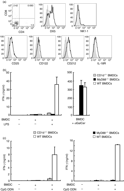
Figure 4.
Invariant natural killer T (iNKT) cell lines express cytokine receptors and become activated via a cytokine-driven mechanism. (a) Expression of surface molecules on iNKT cells. iNKT cell lines were stained with anti-CD3e-fluorescein isothiocyanate (FITC), CD1d-tetramer-allophycocyanin, and a panel of surface molecules. Expression of CD4, CD8, DX5, NK1.1, CD25, CD122, CD212 and interleukin (IL)-18R was determined on CD3-positive CD1d-tetramer-positive NKT cells. The open histograms indicate staining with each monoclonal antibody (mAb) and the grey histograms represent the isotype controls. Results are representative of two separate experiments. (b) iNKT cell line activation by lipopolysaccharide (LPS)-stimulated bone marrow dendritic cells (BMDCs) is CD1d and MyD88 dependent. 1 × 10⁵ NKT cells were incubated with LPS (10 μg/ml) or α-galactosylceramide (αGalCer; 10 ng/ml) in the presence of 1 × 10⁵ wild-type (WT) BMDCs, CD1d−/− BMDCs, or MyD88−/− BMDCs. Supernatants were collected and interferon (IFN)-γ production was determined by enzyme-linked immunosorbent assay (ELISA). (c) CD1d- and MyD88-dependent iNKT cell line activation by CpG oligodeoxynucleotide (ODN). 1 × 10⁵ NKT cells were incubated with CpG ODN (125 ng/ml) in the presence of 1 × 10⁵ WT BMDCs, CD1d−/− BMDCs or MyD88−/− BMDCs. IFN-γ production in supernatants was measured by ELISA. Results are representative of two separate experiments.

