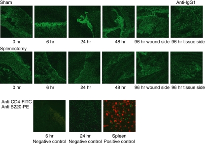
Figure 5.
The deposition of antibodies to damaged tissues in wound healing. Two-month-old C57BL/6 mice were splenectomized or sham-operated. One week later, 3-mm punch biopsies were performed on the backs of these mice. Tissue samples were obtained by taking 6-mm punch biopsies around 3-mm holes. Frozen samples were analysed with fluorescein isothiocyanate (FITC) or phycoerythrin (PE)-labelled antibodies and observed by confocal microscopy. The deposition of antibodies to damaged tissues was analysed by FITC-labelled anti-mouse immunoglobulin G1 (IgG1). Wounded tissues were stained by FITC-labelled anti-mouse CD4 (T cells) and PE-labelled anti-mouse B220 (B cells). Neither T nor B cells were observed, which became negative controls to FITC-labelled anti-mouse IgG1. As a positive control, spleens were stained with anti-mouse CD4 (T cells) and PE-labelled anti-mouse B220 (B cells).

