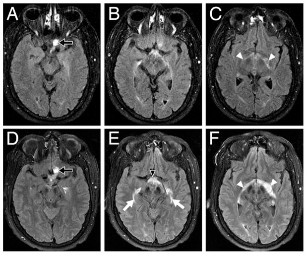
Fig. 2.
Axial T1-weighted FLAIR MR images demonstrating the progression of hemangioblastoma-induced peritumoral edema involving the optic pathways from August 2005 to January 2007. In August 2005 (A, B, and C), edema surrounding the left optic nerve hemangioblastoma (A, arrow) with mild edema (C, arrowheads) was noted along the optic fibers bilaterally. In January 2007 (D, E, and F), 5 days before surgery, the size of the hemangioblastoma had remained stable (D, arrow), but a significant increase in edema in the left optic nerve extended through the optic chiasm (E, arrowhead) and lateral geniculate bodies (F, arrowheads) and into the bilateral optic radiations (E, arrows).

