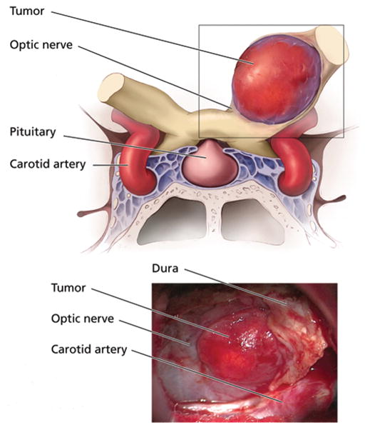
Fig. 3.

Upper: Coronal illustration showing the relationship of the hemangioblastoma to the optic apparatus and surrounding anatomical structures. Lower: Intraoperative photograph showing the vascular hemangioblastoma arising from within the left optic nerve and separating the fibers of the medioinferior aspect of the nerve.
