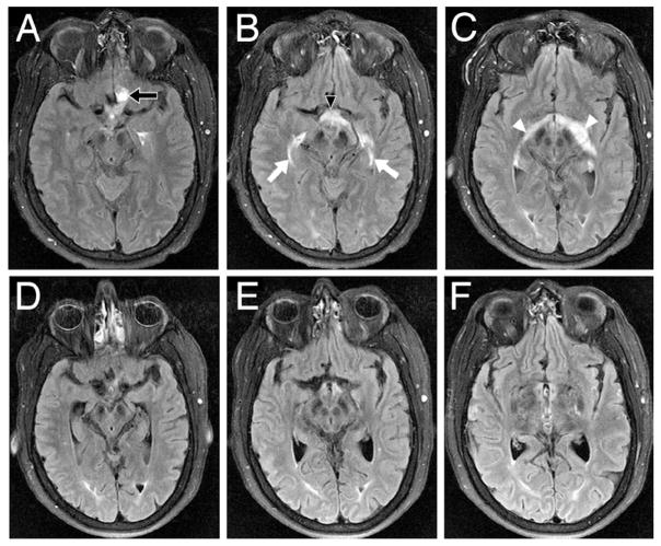
Fig. 4.
Axial FLAIR MR images (A, B, and C) obtained preoperatively, revealing significant edema surrounding the left optic nerve hemangioblastoma (A, arrow) that tracked into the optic chiasm (B, arrowhead) and along the bilateral optic system (C, arrows and arrowheads). Axial FLAIR MR images (D, E, and F) obtained 7 days after surgery, demonstrating resolution of edema.

