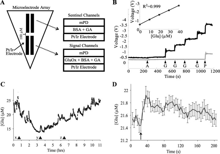
Figure 1.
In vitro calibration and in vivo assessment of microelectrode arrays. A, Schematic of the self-referencing microelectrode arrays. Signal channels are coated with GluOx and are responsive to glutamate, whereas sentinel channels are coated only with the cross-linking proteins BSA and glutaraldehyde (GA). Signal and sentinel channels both have an m-phenylenediamine exclusion layer (mPD). B, Typical in vitro calibration of signal (black line) and sentinel (gray line) channels. Arrows indicate the time of application of ascorbic acid (A; 500 μm), glutamate (G;10 μm), and peroxide (P). Both channels show little or no response to ascorbic acid but respond robustly to peroxide, whereas only the signal channel responds robustly to glutamate. Inset, Calibration curve demonstrating the linear response of the signal channels to changes in glutamate concentration. C, Changes in glutamate concentration after pentobarbital injection in one rat (x; 50 mg/kg, i.p.). The first 90 s episode of arousal with locomotion was observed at (y), but full motor activity was regained only at (z). D, Rapid increase in glutamate concentration during whisker stimulation contralateral to an electrode implanted in the barrel cortex in one rat. The rat received 15 stimulations applied over the course of 90 min. The average of all 15 stimulations, time locked to stimulation onset, is depicted in 4 s intervals. Stimulation onset began at arrow and each stimulation lasted on average 23.72 ± 3.28 s.

