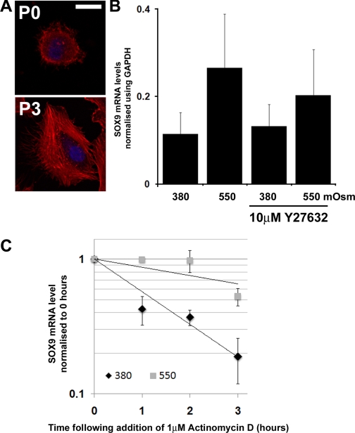
Fig. 2.
Effect of hyperosmolarity on SOX9 mRNA levels in freshly isolated HAC. A: fluorescence micrographs showing distribution of actin (stained with rhodamine-conjugated phalloidin-red) in HAC when they are freshly isolated (P0) or after three passages in monolayer culture (P3). Cell nuclei are stained with DAPI (blue). Images are representative of the majority of cells in the cultures. Scale bar = 10 μM. B: real-time PCR analysis of SOX9 mRNA levels in freshly isolated HAC cultured at 380 or 550 mosM in the presence and absence of Y27632 (10 μM) for 5 h. Data represent means and SDs obtained from experiments with cells from 4 different donors. C: SOX9 mRNA decay in freshly isolated HAC cultured at different osmolarities. Within 48 h of extraction from tissue, HAC were cultured at 380 or 550 mosM for 2 h before addition of actinomycin D. RNA was then extracted in triplicate at 0, 1, 2, and 3 h for reverse transcription and analyzed by real-time PCR. Data represent means and SDs of the fold changes in SOX9 mRNA levels compared with time point 0 (n = 4).

