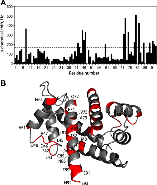
Fig. 11.
S100A1 binds directly to PKA. A: chemical shift perturbations are illustrated for Ca2+-S100A1 on binding the PKA regulatory subunit peptide. B: chemical shift changes mapped on the Ca2+-S100A1 structure. Regions of Ca2+-S100A1 that have large chemical shift, defined as Δδ > 170 Hz, are colored in red. The majority of these residues are located in the hinge region (residues 40–50) and helix 4 (residues 72–89). These regions are close together in three-dimensional space and comprise a large part of the hydrophobic protein binding cleft of Ca2+-bound S100A1.

