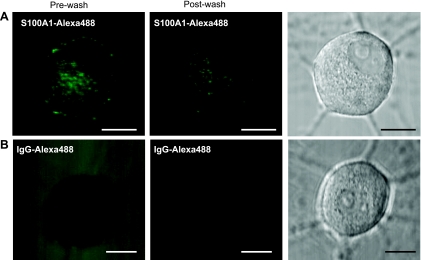
Fig. 2.
Extracellular S100A1 is taken up by SCG neurons. Representative single-plane confocal images of neurons treated for 24 h with S100A1-Alexa 488 (5 μM; A) or with Alexa 488-IgG (20 μg/ml; B) before (left) and after (center) washout of the extracellular media. Right: corresponding transmitted light images. Scale bars, 10 μm.

