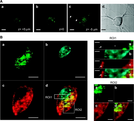
Fig. 3.
Intracellular distribution of exogenous S100A1. A: confocal x–y images at different z-axial planes (a, top; b, middle; c, bottom) from a neuron treated with extracellular S100A1-Alexa 488 (10 μM) for 24 h. S100A1-Alexa 488 fluorescence is located in the cytoplasm and present in the neuronal processes (arrows in c) but not in the nucleus. Scale bars, 10 μm. B: representative confocal images of another neuron treated with extracellular S100A1-Alexa 488 for 24 h (a) and colabeled with DAPI (b) to stain the nucleus and FM4-64 (c) to stain plasma membrane and to follow bulk membrane internalization; d shows the merge of a–c. ROI1 and ROI2 are enlarged views of the outlined regions of interest in Bd, demonstrating clear S100A1 exclusion from the nucleus and association with membranous structures.

