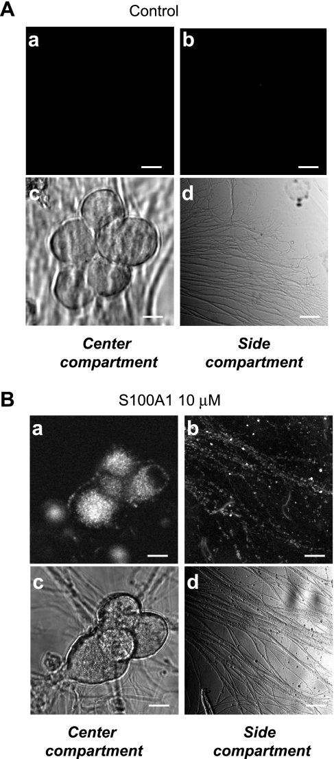
Fig. 5.
Extracellular S100A1 is taken up and undergoes retrograde axonal transport. Representative confocal (a and b) and transmitted light (c and d) images of center and side compartments of control (A) and S100A1-treated (B) compartmented SCG cultures. Only the side compartments shown in A and B were treated with Alexa 488-IgG or S100A1-Alexa 488 (5 μM), respectively. After 24 h chambers were rinsed and imaged. S100A1-Alexa 488 fluorescent signal was present in axons in the side compartment and in axons and cell bodies of the central compartment, indicating uptake and translocation of S100A1 from axonal projections toward somas via retrograde transport. No uptake or translocation was seen in Alexa 488-IgG treated neurons (A). Scale bars, 10 μm.

