Fig. 6.
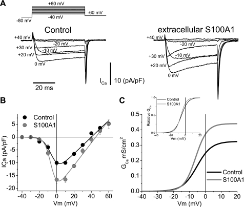
Extracellular S100A1 enhances whole cell Ca2+ currents in SCG neurons. A: representative macroscopic Ca2+ current traces from control (left) and S100A1-treated (10 μM; right) neurons. Macroscopic currents were elicited by depolarizing pulses with the voltage protocol illustrated at top, with 2 mM Ca2+ as the charge carrier. B: average peak current density plotted against voltage (I-V) relationship for step depolarizations from control (black circles) and S100A1-treated (10 μM; gray circles) neurons. The solid lines through the I-V curves in B are least squares fits of the data to a modified Boltzmann-ohmic equation. Average parameter values for maximum conductance (Gmax), half-activation potential (Vh), and steepness (k) for control neurons were 0.32 mS/cm2, −5.3 mV, and 4.5 mV, respectively. For S100A1-treated neurons Gmax, Vh, and k were 0.43 mS/cm2, −6.1 mV, and 3.6 mV, respectively. Vm, membrane potential. C: conductance (GCa) vs. V relationship derived from I-V plots from control and S100A1-treated neurons. Inset: normalized G vs. V plots from C.

